Who this is for
- You were told an X-ray shows a “cyst,” “granuloma,” “lesion,” “shadow,” or “infection” near a tooth root and want to know what that means.
- You have a tooth that was root-canal treated years ago and the dark area is still there or has grown.
- You have swelling, a draining pimple on the gum, numbness, or a tooth that is loosening for no clear reason.
What we do at our office
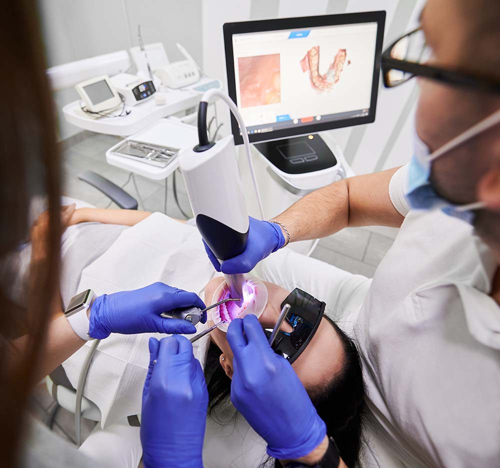
Dr. Baruch Tetri, DDS, is a periodontist, a gum and jawbone specialist, and reading the bone around teeth is his daily work. He tests whether the nearby teeth are alive, compares the X-ray with the examination, and names the finding by what it is doing rather than by its shape. Most dark areas at a root tip are inflammation from a dead nerve: a granuloma (inflamed tissue, nothing to do with the disease of the same name) or a radicular cyst (a fluid-filled sac that grows from it). An X-ray cannot tell the two apart; only tissue under a microscope can, and usually it does not need to, because both usually heal after root canal treatment.
What happens at the visit
- Reading the image. We look at your existing X-rays and take new ones only if the question needs them.
- Testing the teeth. Cold and tapping tests on the suspect tooth and its neighbors, gum probing, a check for cracks and old fillings. One silent tooth or one dark area is not enough on its own.
- A 3D scan when it changes the plan. A limited CBCT (a three-dimensional dental X-ray) shows how far the area reaches, whether it touches the sinus or a nerve, and whether an old root filling is short.
- Treatment. We treat the cause we found: most inflammatory lesions start to heal once the infected canal is cleaned and sealed. A previously treated tooth may need retreatment first.
- Surgery when it persists. If the area stays or grows after a sound root canal, the root tip and the lesion are removed through a small opening in the gum, and the tissue goes to a pathology lab.
- Follow-up. We compare X-rays at intervals over months; bone fills in slowly, and comfort returns before the image catches up.

Risks and limits
- Not every dark area is infection. Rarely it is something else, which is why we check the image against the tooth and widen the search when they disagree.
- A lesion can persist after a technically sound root canal; that calls for re-diagnosis, not an automatic repeat.
- Surgery near the sinus or the nerve of the lower jaw carries a small risk of sinus involvement or altered sensation; we map these on the scan first.
- A tooth that cannot be restored is removed; the socket is then planned with any replacement in mind.
- A lesion that suddenly swells or starts to drain means call us the same day.
Questions patients ask
Does a cyst mean surgery?
Usually not. If the cause is a dead nerve, a root canal removes it and many heal afterward over months. Surgery is for lesions that persist after that, are very large, or do not behave like infection.
Will antibiotics shrink it?
No. Antibiotics cannot reach the source inside the tooth and do not tell a cyst from a granuloma; they are used only when infection is spreading.
Can the area come back after treatment?
It can, which is why we follow it with comparable X-rays until the bone has filled in. A recurrence is re-diagnosed before anything is repeated.
How long until it is gone on the X-ray?
Months, often longer; symptoms settle first. We tell you the review interval and what would bring you in sooner.
Sources
- ESE S3 guideline: Treatment of pulpal and apical disease
- AAE/AAOMR: CBCT in endodontics position statement
- PubMed: Clinical versus histopathologic diagnosis of periapical lesions
- PubMed: CBCT accuracy for cyst versus granuloma
- PubMed: Non-endodontic lesions mimicking apical disease
Request a visit
Tetri’s Smile · 16690 Collins Avenue, Suite 701, Sunny Isles Beach, FL 33160 · Serving Aventura, Bal Harbour, North Miami Beach, Hallandale Beach and greater Miami.