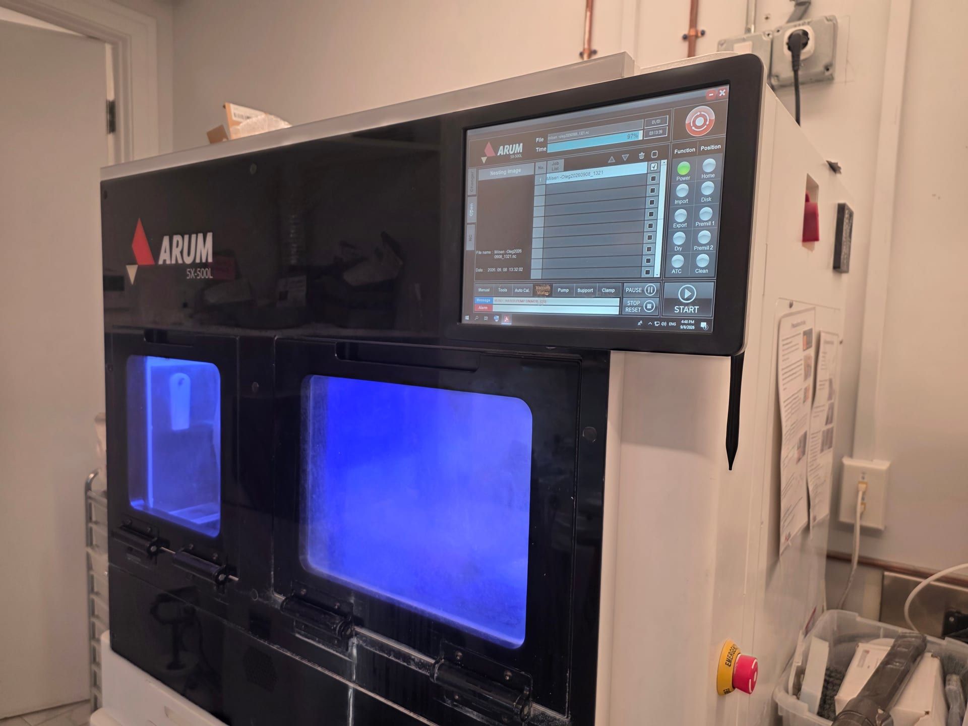
3D imaging: Planmeca Viso G7
The Viso G7 is a CBCT scanner (cone-beam computed tomography, a three-dimensional dental X-ray). In plain terms, Planmeca documents that it can image a single tooth in fine detail, for root canal work, or the whole jaw and the jaw joints, at a low dose, and that it corrects for small movements so a retake is rarely needed. The scan is sized to the question: we choose the field, up to 30 × 30 cm, and the protocol for the anatomy and the diagnostic question.
Review and planning: Planmeca Romexis
Romexis is the software that reads the scan. We review the volume in several planes (front to back, side to side and from above) and alongside your photographs and digital impressions, so the diagnosis and the surgical or restorative plan come from one set of records.
Jaw-function records: K7, surface EMG and TENS
When the examination calls for it, three records can be added. K7 jaw tracking uses a small magnet and sensors to record how the lower jaw moves. Surface EMG (electromyography) uses skin sensors to record how hard the jaw muscles are working. TENS (transcutaneous electrical nerve stimulation) is a mild electrical stimulation of the jaw muscles, used during a functional evaluation. They help us ask better questions; they do not decide treatment. Reversible care, such as a bite appliance (a removable splint worn over the teeth), comes before any irreversible dental work.
Implant position capture: PIC photogrammetry
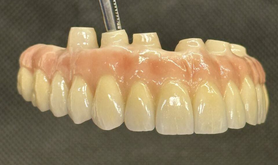
For selected complete-arch implant cases (a full arch of teeth on several implants), PIC photogrammetry uses cameras to record the positions of small markers placed on the implants and captures the relative positions of the implant components for the digital restorative workflow. Accuracy depends on the system, its calibration, how the images are taken and the conditions of the case.
Design and fabrication: the laboratory in the building
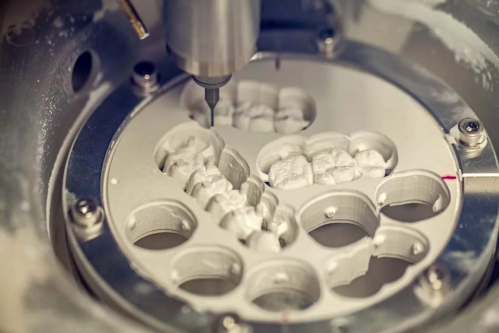
Our clinical and laboratory teams work from the same digital model, the proposed bite, the chosen materials and the approved design. Design and milling happen in the building; materials and timing depend on the case. A trial stage, such as a provisional (a temporary tooth or bridge made to the planned shape) or a bite appliance, lets us check the plan in your mouth before anything final is made.
How the records become a plan
- Ask the clinical question. The examination determines which record could change the plan.
- Capture only what is useful. A focused or a larger field is chosen by the anatomy, the diagnosis and the lowest dose that answers the question.
- Align the records. Imaging, photographs, digital impressions, functional data and your history are reviewed as one case.
- Test the plan. A provisional or a bite appliance goes in first, and we adjust it before the final work is made.
- Design and fabricate. The laboratory works from the approved clinical position, the material plan and the goals of your case.
- Verify and maintain. Fit, function, how the gums respond, home care and follow-up remain part of the result.
Questions patients ask
Will I need a 3D scan?
Only when the examination raises a question a regular X-ray cannot answer, such as where a nerve runs before implant or wisdom-tooth surgery.
Can you use scans I already have?
Often, yes, if they are recent and cover the right area; ask the front desk how to send them.
Does the jaw-tracking equipment diagnose TMJ problems?
No. TMJ (jaw joint) disorders are diagnosed from your history and the examination; the records add detail.